More at the artist's web site, and here's a mini documentary on the poultry portraiture project.
Amazon Deals
New at Amazon
Wednesday, May 3, 2023
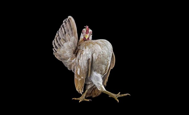
May 4th is International Respect for Chickens day: some chicken-related links to help you celebrate
More at the artist's web site, and here's a mini documentary on the poultry portraiture project.
Thursday, March 2, 2023
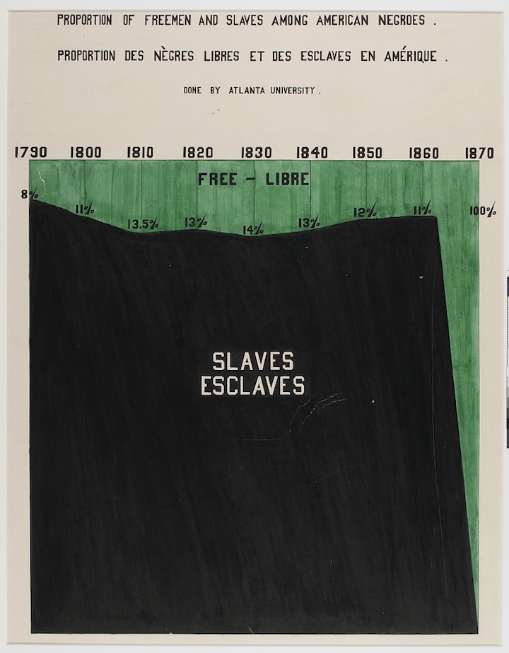
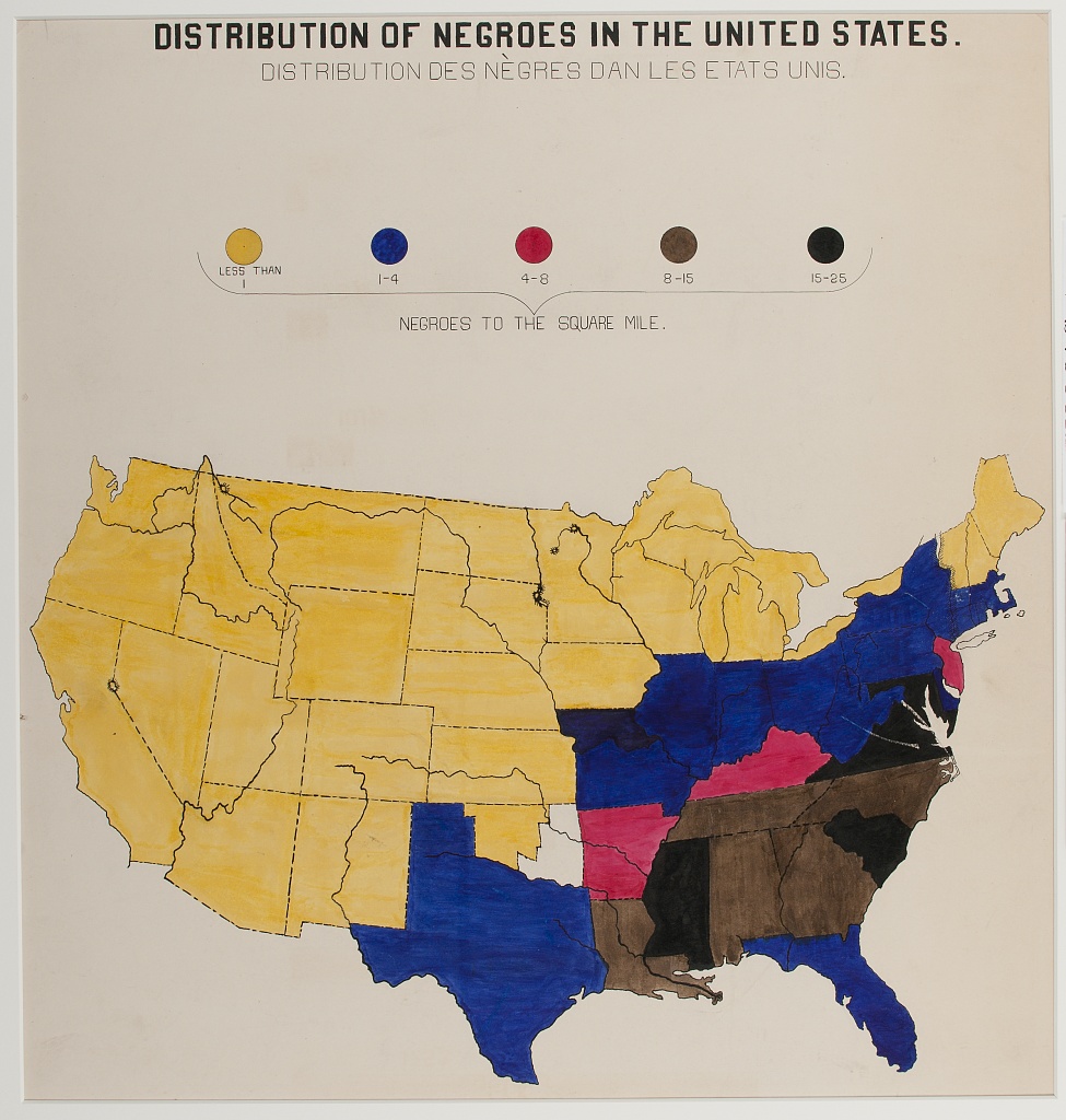
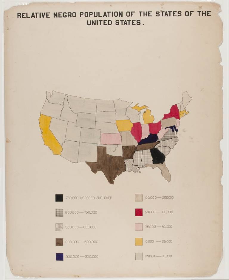
W. E. B. Du Bois’s hand-drawn infographics describing black life In 1900
Via Flashbak:
Friday, February 17, 2023
How to blow a cow
Cow blowing, per Wikipedia:
Kuhblasen, phooka, or doom dev, is a process used in many countries according to ethnographers, in which forceful blowing of air into a cow's vagina (or sometimes anus) is applied to induce her to produce more milk.
Cow blowing was the reason why Gandhi abjured cow milk, saying that "since I had come to know that the cow and the buffalo were subjected to the process of phooka, I had conceived a strong disgust for milk."
Thursday, February 16, 2023
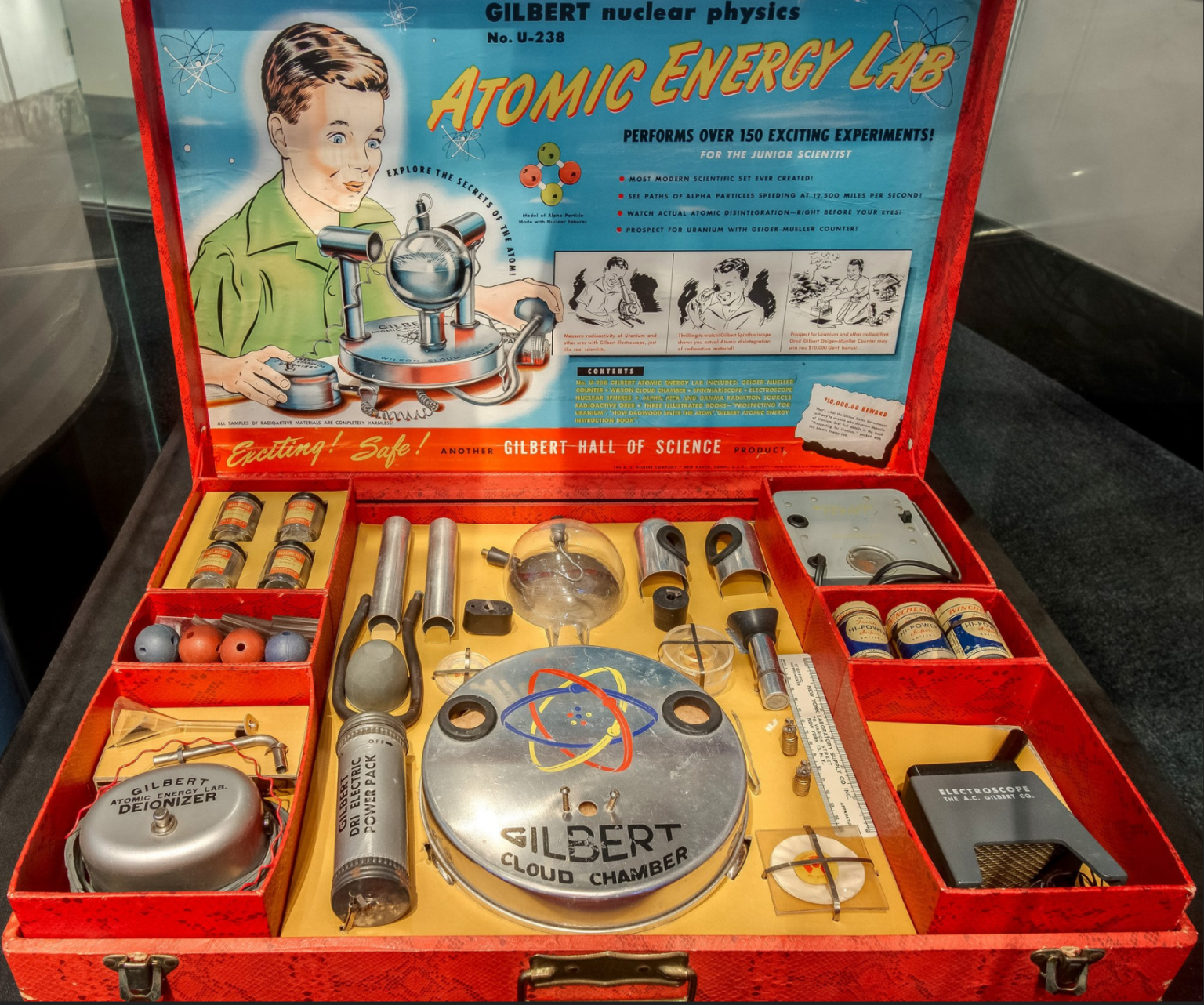
Toys from the past - Radioactive Atomic Energy Lab Kit with Uranium (1950)
![]() |
| Radioactive Atomic Energy Lab Kit with Uranium (1950) |
Here's an Atlas Obscura video with more:
Thursday, February 2, 2023
Study Result
Impression
IMPRESSION:
1. Stable postoperative changes of the stomach and bowel without evidence of obstruction.
2. Hepatic lesions appear slightly increased in size and are again concerning for metastatic disease.
3. Interval decreased ascites and resolved pneumoperitoneum. Resolved bilateral pleural effusions.
4. Mild wall thickening of the bladder with punctate foci of intraluminal gas may reflect recent instrumentation versus cystitis.
5. Mild ascending aortic aneurysm measuring 4.1 cm.
Images and interpretation personally reviewed by: Sudeep Soni, MD
Narrative
EXAM: CT CHEST/ABDOMEN/PELVIS W/ IV CONTRAST
INDICATION: Primary colon cancer, also with history of renal cell and prostate cancer.
TECHNIQUE: CT of the chest, abdomen, and pelvis with intravenous contrast was performed. Multiplanar reconstructions were performed.
COMPARISON: CT abdomen and pelvis 9/10/2022, CT chest abdomen pelvis 8/31/2022
FINDINGS:
CHEST:
Lung/Airways: Central airways are patent. No consolidation. No suspicious lung nodule.
Pleura: No pleural effusion or pneumothorax.
Thyroid: Stable subcentimeter right thyroid nodule.
Mediastinum: No mediastinal mass. Esophagus is normal.
Lymph nodes: No thoracic adenopathy.
Heart: Heart size is normal. Coronary artery calcifications. No pericardial effusion.
Vasculature: Mildly dilated ascending thoracic aorta measuring 4.1 cm, previously 3.9 cm. Normal caliber pulmonary trunk. Right IJ chest port with catheter tip at the superior cavoatrial junction.
Subcutaneous Tissues/Chest Wall: No concerning soft tissue mass.
ABDOMEN:
Liver: Redemonstrated multiple hepatic lesions. Reference posterior right hepatic lobe lesion measures 2.5 x 2.9 cm (2/92), previously 2.7 x 2.2 cm. Segment 8 lesion measures 1.8 x 1.7 cm (2/89), previously 1.7 x 1.4 cm.
Hepatic vein(s): Patent.
Portal vein(s): Patent.
Gallbladder/Bile Ducts: Cholelithiasis with nonspecific pericholecystic fluid and setting of ascites. No biliary ductal dilatation.
Spleen: Normal size.
Pancreas: No pancreatic mass or pancreatitis. Pancreatic duct normal in caliber.
Adrenal Glands: No adrenal nodule.
Kidneys: Stable small renal hypodensities. No hydronephrosis or nephrolithiasis.
Bowel: Stable postoperative changes of the stomach and bowel without evidence of obstruction. Redemonstrated fluid-filled bowel loops with air-fluid levels.
Peritoneum/Retroperitoneum: Interval decreased ascites. Previously seen pneumoperitoneum has resolved.
Lymph Nodes: No abdominal adenopathy.
Vasculature: Normal caliber abdominal aorta and iliac arteries. Mild scattered atherosclerotic disease.
PELVIS:
Reproductive Organs: Status post prostatectomy.
Bladder: Mild circumferential wall thickening with foci of intraluminal gas.
Lymph Nodes: No pelvic lymphadenopathy.
Vasculature: Unremarkable.
Subcutaneous Tissues/Abdominal Wall: No hernia.
SKELETAL STRUCTURES: No acute fracture or suspicious osseous lesion. Partially imaged left hip arthroplasty. Bilateral glenohumeral osteoarthritis, right greater than left.
Component Results
There is no component information for this result.
General Information
Ordered by Shruti Murali
Collected on 01/27/2023 9:08 AM
Resulted on 01/27/2023 9:42 AM
Result Status: Final result